
Research Activities
CREATIS, CNRS UMR 5220 – Inserm U1294, Myriad team
Multimodal generative AI for medical imaging
Deep learning and image processing focused on segmentation,
image analysis and synthetic data generation
Research Domains
- Since 2023 : CREATIS Laboratory, Myriad team
- Generative AI, Latent Diffusion Models conditioned by textual prompt, segmentation and classification using Deep Learning, instance segmentation, brain imaging, multiple sclerosis lesion segmentation and longitudinal follow-up, biomedical and medical imaging.
- 2015-2023: CITI Laboratory
- Swarm Robotics, Software-Defined Radio, Physical Layer of Wireless Communications, Deep Learning applied to communication systems.
- 2000-2013: CREATIS Laboratory
- Medical Image Processing, Segmentation, Region Growing, Multidimensional Approach, Classification in Feature Space, Mean-Shift, Variational segmentation, Integration of shape prior, Medial Representations, and Statistical Shape Model, Bone architecture and osteoporosis.
Current Research Directions
- 1. Multimodal generative AI for brain MRI (Dall-e Brain project)
- Healthy-subject data are scarce in brain imaging. This direction develops text-conditioned generative models able to synthesise realistic 2D MRI slices (T1-w, T2-w, FLAIR) from anatomical and clinical textual descriptions. It relies on an original corpus of 2 million image-text pairs, automatically built from four public databases (IBSR, OASIS, KIRBY, IXI) by registration on anatomical atlases, and on Latent Diffusion Models. The approach opens perspectives for rare data augmentation, for modelling the temporal evolution of chronic diseases, and for the identification of latent biomarkers. Code is released as open source on GitLab. Computations are carried out on the Jean Zay supercomputer (IDRIS/CNRS), with an allocation renewed in February 2026 (H100 and V100 hours).
- 2. Segmentation and longitudinal follow-up of multiple sclerosis lesions (TIME project)
- Detecting lesions that appear between two successive examinations is fundamentally different from segmenting a single time point: analysing each examination independently fails to preserve temporal consistency. This direction addresses two complementary obstacles. Cross-site generalisation is studied through a systematic evaluation of the transferability of nnU-Net across three databases with very different characteristics (MSSEG-1, MSLesSeg, OpenMS), quantifying the specialisation/generalisation trade-off. Longitudinal segmentation is addressed by LAS-Net (Longitudinal Attention Segmentation Network), a transformer architecture based on SwinUNETR whose attention windows are calibrated to the characteristic size of MS lesions, which reaches an accuracy close to human expertise for the detection of very small new lesions. This work is conducted within the MUSIC transversal project of CREATIS and in collaboration with ENET'Com (Tunisia).
- 3. Deep Learning for the segmentation of biological and physico-chemical structures
- In collaboration with the BF2I laboratory (Functional Biology, Insects and Interactions), deep learning methods are applied to the automatic segmentation of bacteriocytes — the cells hosting symbiotic bacteria in aphids — from bright-field microscopy images, comparing Mask R-CNN and YOLO. With the IRCE laboratory (Institut de Recherches sur la Catalyse et l'Environnement), the same approaches are applied to the segmentation of nanocrystals and water droplets during in situ condensation observed by environmental transmission electron microscopy.
- Convergence of the two main directions
- These directions are not independent. A latent diffusion model trained jointly on healthy and pathological MRI encodes in its latent space a regular representation of normal and lesional brain anatomy. This representation can be exploited as an anatomical prior to constrain the longitudinal segmentation model: formulated in the latent space rather than in the noisy image space, new-lesion detection becomes better conditioned and more robust to domain shift. This convergence is the central object of Maël Rocher's doctoral thesis.
Collaborations and Networks
- International
- University of North Carolina at Chapel Hill, USA
(2011-2026): long-standing collaboration with Juan Carlos Prieto, formerly a
co-supervised doctoral student and now Research Assistant Professor, on medial
representations and, more recently, on multimodal generative AI.
ENET'Com, University of Sfax, Tunisia (2024-2026): active collaboration with F. Kallel and M. Sahnoun on multiple sclerosis lesion segmentation. - National and multidisciplinary
- BF2I laboratory, Lyon (since 2024) on the automatic
segmentation of bacteriocytes.
IRCE laboratory, Lyon (since 2023) on the segmentation of nanocrystals in electron microscopy.
MUSIC transversal project of CREATIS (Multiple Sclerosis and neuroinflammation, 15 members): active member since 2023, co-director since 2026 jointly with F. Durand-Dubief, neurologist at the Hospices Civils de Lyon. - Research networks
- Member of LabEx PRIMES (Physics, Radiobiology, Medical
Imaging and Simulation) since 2012, contributing to working groups WP4
(multidimensional data processing) and WP5 (imaging and simulation).
Participation in the CNRS GDR ISIS/IASIS (theme B, Image and Vision) and CNRS Inserm GDR STIC-Santé (theme B, Signals and Images in Health).
Scientific Community Service
- Reviewing
- Neurocomputing (Elsevier, 2025); IABM 2026, the French colloquium on Artificial Intelligence in Biomedical Imaging.
- Doctoral juries and committees
- Reviewer (rapporteur) of the thesis of Sayeh Gholipour
Picha, GIPSA-Lab, Université Grenoble Alpes, defended 16/12/2025.
Jury member for the thesis of Radu Dragos Urs, Université Bordeaux 1, defended 29/03/2013.
Member of the individual monitoring committee of Aya Jendoubi's thesis, École Centrale de Lyon, since 2025, as representative of the EEA doctoral school. - Recruitment and organisation
- Member of the selection committee for an Associate
Professor position in CNU section 61 at ENIB, Brest (2025).
Member of the organising committees of the DLMI 2025 school (Deep Learning for Medical Imaging) and of the IABM 2026 colloquium. - Technology transfer
- Two expert consultancy contracts with Bovo Predict (2025-2026, as co-holder), a startup developing AI solutions for dental imaging, on machine learning methods for the prediction of bone loss ahead of tooth extraction.
- Outreach
- Contribution to the L'Oréal Foundation programme "For Girls in Science", speaking to high-school students (February 2025).

Dall-e Brain — text-conditioned brain MRI generation (EUSIPCO 2025)
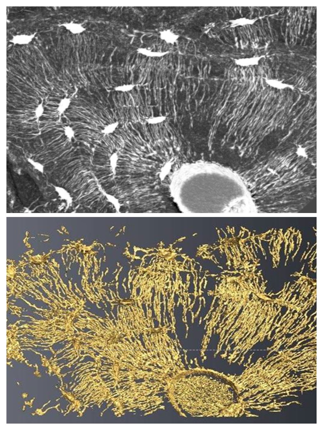
SRµCT images of bone tissue cellular structure - FRM Microtomos national project.
- Energy derived from the Sato filter
- Improved extraction of tubular structures
- Enhanced connectivity of the canaliculi