Metabolism Disorders
Thanks to the concept of integrated multiparametric MR imaging (IMPMRI) previously introduced, we envision a multiorgans and multiparametric approach to identify new biomarkers of (i) Non-Alcoholic Steatohepatitis (NASH) in patients with NAFLD or (ii) to monitor experimental protocols such as overfeeding or fasting related to nutrition. In the liver, IMPMRI will be extended to the quantification of collagen and short T2 species quantity with new generation of MR sequence using spiral k-space encoding. The large amount of quantitative information can be also associated with radiomics and mined approaches to develop models of prediction of major outcomes in metabolic diseases.
Keywords: Multiparametric MRI, Quantitative MRI, Biomarkers, NAFLD, NASH, Fasting, Overfeeding , Radiomics
Radiomics in Oncology
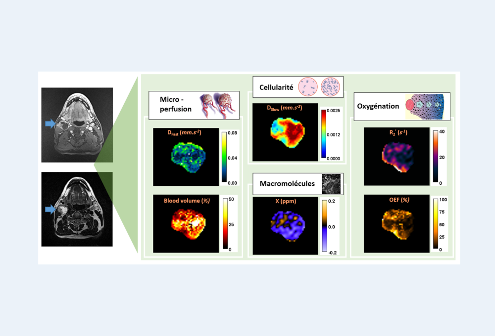
Thanks to the concept of integrated multiparametric MR imaging (IMPMRI) previously introduced, a multiparametric acquisition protocol was integrated in breast imaging clinical workflow at the comprehensive cancer center Léon Berard. The extension of IMPMRI concept will allow the reconstruction of orthogonal, quantitative and multiparametric maps probing tumor biology at the macroscopic scale. These maps constitute the source of radiomics containing tumor phenotypic information at different levels. Artificial intelligence will be used to mine these data and provide predictive information for a personalized diagnosis but also to speed-up parametric maps reconstruction.
In this axe, we also interest about:
- Rare and pediatric tumors such as Ewing Sarcoma and Osteosarcomas, particularly in the development of predictive models of neo-adjuvant chemotherapy histological response and survival using radiomics ,
- Lung tumors stratification and response to immunotherapy using radiogenomics in partnership with the Center of cancer research of Lyon (CRCL).
Keywords: Multiparametric MRI, Quantitative MRI, Biomarkers, Precision medicine, Breast cancer, Rare and Pediatric tumors, Radiomics, Radiogenomics
Magnetic Resonance Elastography
Magnetic Resonance Elastography (MRE) aims at producing a quantitative cartography of the viscoelastic properties of tissues which is an interesting biomarker of the evolution of various pathologies. Ongoing work focus on all three aspects of MRE experiments, namely
- dedicated instrumental setups for ex vivo and in vivo measurements which integrate shear wave generation as well as auxiliary functions
- specific MRE sequences which include multifrequency strategies and optimal control approaches coupled with rapid detection schemes such as UTE.
- advanced reconstruction algorithms
In vivo applications target liver and brain MRE.
Multiple Sclerosis and Neuroinflammatory diseases
Magnetic resonance imaging (MRI) has become a cornerstone in monitoring disease activity and progression in patients diagnosed with multiple sclerosis (MS) as well as associated inflammatory diseases such as neuromyelitis optica spectrum disorders (NMOSD).
In MS, common progression markers include lesion and atrophy measures with a focus on T2 lesion load. However, these markers are moderately correlated with the clinical status of the patient and do not allow the prediction of the disease progression. Thus, advanced MR techniques such as diffusion MRI (DTI), resting-state fMRI, spectroscopic imaging (MRSI), and magnetization transfer (MT) are developed in view of measuring and modeling brain connectivity based on graph theory. Morphology (Thickness of Gray Matter), WM micro-architecture (diffusion MRI providing structural connectivity) and GM function (Resting-state fMRI) through the analysis of several network metrics of integration, segregation or hubs are providing a sensitive characterization of alterations in brain connectivity in relation with the inflammatory and neurodegenerative processes occurring in MS patients. Based on these initial measures and analysis of the different brain connectivity, machine and deep learning processing, mainly based on Convolutional Neural Networks (CNN), and Graph CNN (GCNN), are developed for the classification of MS patients (CIS, RR, SP & PP), and for the prediction of the disease course and the patient status. This work is based on the OFSEP project coordinated in Lyon (S. Vukusic).
Neuromyelitis optica spectrum disorders (NMOSD) has been recently introduced as ab-mediated demyelinating disorders where patients commonly have serum antibodies against aquaporin-4 (AQP4) protein as well as MOG-Ab associated disease, where patients have serum antibodies against myelin oligodendrocyte glycoprotein. NMOSD patients mainly suffer from optic neuritis and/or longitudinally extensive myelitis (LETM), which attacks lead mainly to spinal cord (SC) atrophy, causing numerous body dysfunctions. However, very little quantitative information has been gathered, especially by imaging, for diagnostics or prognosis in NMOSD. Our project endeavours to further characterise and differentiate the SC damages in the two antibody subgroups (AQP4 & MOG) associated with NMOSD, in comparison with clinically isolated syndrome (CIS) patients and healthy controls, by implementing sensitive and clinically applicable in vivo MRI biomarkers. DTI and PSIR sequences are developed for measuring WM changes and tissue atrophy during the disease evolution. This work is based on the NOMADMUS project in Lyon and collaborations with Berlin group (F. Paul).


